Case Study
Anca Chiriac, Florin Maghiar, Uwe Wollina
Pityriasis rosea bei älteren Patienten – eine Fallserie
Pityriasis rosea of the elderly – a case series
(NACH CARE-LEITLINIE)
Keywords | Summary | Correspondence | Literature
Keywords
elderly, herald patch, Pityriasis rosea, scaling
Schlüsselworte
ältere Patienten, Pityriasis rosea, Primärmedaillon, Schuppung
Summary
Pityriasis rosea (PR) is an inflammatory self-limiting dermatosis common in younger ages, whereas it is rather uncommon in patients 65+. We report a series of 7 patients (65 to 75 years old, 2 males and 5 females) with PR. The clinical presentation was peculiar because of widespread distribution with rather erythematous, inflammatory lesions but asymptomatic. There was no association to common viral disorders including H1N1 or COVID-19 nor to vaccination against. No drug-induced PR was noted. No treatment was necessary, and the course was mild. PR of elderly is uncommon but has some peculiarities. Treatment was unnecessary.
Zusammenfassung
Die Pityriasis rosea (PR) ist eine entzündliche, selbst-limitierte Dermatose, die vor allem bei jüngeren Patienten vorkommt. Bei Menschen über 65 Jahren ist sie rar. Wir berichten über 7 Patienten im Alter von 65 bis 75 Jahren, 2 Männer und 5 Frauen, mit PR. Das klinische Bild zeigte Besonderheiten mit großflächiger, disseminierter Ausbreitung, stärker entzündlichen, erythematösen Läsionen, jedoch ohne Juckreiz. Es fand sich weder eine Assoziation zu häufigen viralen Infekten einschließlich H1N1-Influenza oder COVID-19 noch zur Vakzination gegen diese. Bei den asymptomatischen Erkrankungen war keine Therapie erforderlich, der Verlauf war mild. Die PR älterer Patienten ist eine eher seltene Erkrankung und zeigt Besonderheiten im klinischen Bild. Eine Therapie war nicht erforderlich.
Introduction
Pityriasis rosea is an acute inflammatory skin disease of children and young adults, although rare cases have been reported in adults. The name of the disease remains unchanged since its first description and is based on the rose-colored scale that characterises the skin disease. The disease is also known as pityriasis circinata, roseola annulata, herpes tonsurans maculosus or Gibert disease [1].
It is a papulo-squamous skin disorder, characterized by disseminated patches on trunk and proximal parts of limbs (named „Christmas-tree“ distribution). The disease starts with a larger patch, named herald patch („mother lesion“), followed within days by similar smaller lesions with typical distribution. Each lesion has a typical aspect, an erythematous patch covered by a collarette rose scale.
The skin lesions have a peculiar evolution that represents a clinical argument for the diagnosis. The existence of a prodrome is controversial, it has not been admitted by many patients and may be a coincidental finding. In children and teenagers, it has been reported the existence of a prodrome characterised by airborne respiratory tract or/and digestive symptoms, associated rarely with systemic symptoms, such as arthralgia or fever. However, in elderly no prodrome has been recognized and no correlation with severe form of the skin disease [1, 2].
Herald patch is seen in almost all cases, although its location can vary a lot; can appear on the trunk, but also on the extremities or neck or anywhere on the skin [1]. Herald patch can be associated with burning sensation or pruritus or be completely asymptomatic and even not observed.
Patients seek for medical advice when generalized skin lesions develop within days after herald patch. The skin lesions appear in crops over weeks and last for weeks. The eruption is generalized, number of lesions can vary from a few to many, covering the trunk, abdomen, limbs or can be located on any part of the body. The lesions are bilateral and symmetrically distributed. On the chest, skin lesions follow Langer’s skin tension lines, giving the characteristic appearance of Christmas tree. The lesions can be, as herald patch, completely asymptomatic or associated to pruritus. Pruritus is an associated symptom.
PR is an acute skin disease with a long evolution, sometimes for more than 6 weeks. It is a self-limiting disorder with no residual lesions, a once in a lifetime skin eruption.

Fig. 1: Herald patch on the back.
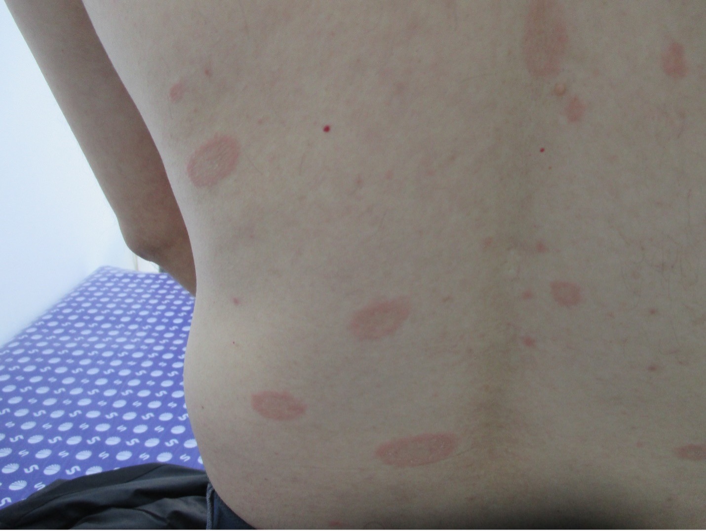
Fig. 2: Christmas tree pattern of pityriasis rosea lesions.
Patients and Methods
During the year 2022, we have seen seven consecutive patients older than 65 years with confirmed PR, but distinct from the classical type. There were 2 males, aged 67 and 70 years, and 5 females, aged 65, 66, 10, 72, 73, and 75 years. Most cases were diagnosed in autumn, with no familial-clustered cases, in patients aged over 70, with slight female predominance. No association with trigger factors has been found. At the time of diagnosis, the patients were in good health state, no viral infection had been detected, no change in medication was reported. All patients were seen before H1N1 vaccination, and they were SARS –CoV-2 negative on polymerase chain reaction. There was no association to COVID-19 vaccination or medical drugs either.

Fig. 3: Rose-colored collarette scaling.

Fig. 4: Multiple erythematous atypical pityriasis rosea lesions
on the lower arm.
Results
Clinical picture comprised of typical lesions of PR spread on large areas of skin not limited to the trunk. Herald patch wad found in most cases, secondary lesions were larger as usual, collarette scales were noted, and the lesions were more inflammatory erythematous (Figs. 1-4). None of these patients suffered from itch.
In many cases, first diagnosis was erythematous scaling skin disorder of unknown etiology in elderly. Despite the impressive clinical picture, the patients were completely asymptomatic but referred to exhaustive investigations for diagnostics.
Treatment was not necessary, since the disease was asymptomatic, and close follow-up confirmed a mild and self-limited course.
Discussion
Diagnosis of PR is clinical, based on the clinical features of the skin lesions, mainly the scale and on the distribution of the lesions. Cutaneous lesions of PR are macules or papules of variable size, ranging from a few mm to many cm, of ovular shape, but can be also round or elliptical or have large, atypical forms. The collarette scale and Herald patch are distinctive and a cornerstone for diagnosis. The scale is rose, attached at the edges and raised at the centre of the lesion. Sometimes the aspect of a typical lesion of PR is coined “cigarette paper”-like due to the scaly feature. No laboratory investigations confirm the diagnosis, but they may be helpful to exclude differential diagnoses.
The real incidence of PR is very difficult to evaluate due to many reasons, such as atypical clinical aspect, the age of patients, misdiagnosis or the link with viral disease. A current cross-sectional study using the All of Us database estimated the overall prevalence as 0.21% [3]. Our case series is unusual since PR has the highest prevalence in the 18 – 25 age group (0.77%). In the age >65, the prevalence drops down to 0.03% [3]. We haven’t seen many cases of PR in this age group in our practise before. This leads to the question of its possible causal relationship.
Unfortunately, the etiology of PR remains unknown, despite it is not a rare skin disease and during COVID-19 pandemic PR-like lesions became part of skin manifestations associated with SARS-CoV-2 infection [4] and COVID-19 vaccination [5-7]. The question about viral etiology of PR still searches for an answer. Many consider PR as viral skin disease, based on seasonal outbreaks of PR, reactivation of human herpes virus 6 and 7 and clinical outcome [8]. But PR-like lesions have reported after many other triggers, such as vaccinations with Bacillus Calmette-Guerin (BCG), H1N1, diphtheria, smallpox, hepatitis B, Pneumococcus. Therefore, PR is considered a para-viral reaction but may occur also after drug intake like captopril, clonidine, barbiturates, clonidine, and imatinib [9]. Although some positive results have been reported for antiviral treatment in the literature [10], most widely used treatment remains symptomatic with topical corticosteroids or calcineurin inhibitors [9, 10].
Korrespondenz-Adresse
Uwe Wollina, MD,
Department of Dermatology and Allergology
Städtisches Klinikum Dresden
Academic Teaching Hospital
Friedrichstrasse 41
DE-01067 Dresden
uwe.wollina@klinikum-dresden.de
ORCID: Uwe Wollina https://orcid.org/0000-0001-5933-2913
Konklusion
In conclusion, our observation is on PR in elderly patients, which is quite uncommon. We found some peculiarities in clinical presentation with widespread eruption of more inflammatory lesions than usual. The disease was not limited to the trunk. Despite this, all patients remained asymptomatic. Treatment was unnecessary and the disease was self-limited. There was no association to SARS-CoV-2 or vaccination for H1N1 or COVID-19. Limitations of this study are the low number of patients and the failure to identify a common cause.
Literatur
1. Villalon-Gomez JM. Pityriasis Rosea: Diagnosis and Treatment. Am Fam Physician. 2018;97(1):38-44.
2. Trayes KP, Savage K, Studdiford JS. Annular lesions: diagnosis and treatment. Am Fam Physician. 2018;98(5):283-291.
3. Joshi TP, Calderara GA, Lipoff JB. Prevalence of pityriasis rosea in the United States: A cross-sectional study using the All of Us database. JAAD Int. 2022; 8:45-46.
4. Wollina U, Karadağ AS, Rowland-Payne C, Chiriac A, Lotti T. Cutaneous signs in COVID-19 patients: A review. Dermatol Ther. 2020;33(5): e13549.
5. Wollina U, Chiriac A, Kocic H, Koch A, Brzezinski P. Cutaneous and hypersensitivity reactions associated with COVID-19 vaccination-a narrative review. Wien Med Wochenschr. 2022;172(3-4):63-69.
6. Temiz SA, Abdelmaksoud A, Wollina U, Kutlu O, Dursun R, Patil A, Lotti T, Goldust M, Vestita M. Cutaneous and Allergic reactions due to COVID-19 vaccinations: A review. J Cosmet Dermatol. 2022;21(1):4-12.
7. Drago F, Ciccarese G, Parodi A. Pityriasis rosea and pityriasis rosea-like eruptions after COVID-19 vaccines. JAAD Int. 2022; 9:127.
8. Leung AKC, Lam JM, Leong KF, Hon KL. Pityriasis rosea: An updated review. Curr Pediatr Rev. 2021;17(3):201-211.
9. Gay JT, Huq M, Gross GP. Herald Patch. In: StatPearls [Internet]. Treasure Island (FL): StatPearls Publishing; 2022.
10. Rodriguez-Zuniga M, Torres N, Garcia-Perdomo H. Effectiveness of acyclovir in the treatment of pityriasis rosea. A systematic review and meta-analysis. An Bras Dermatol. 2018;93(5):686-695.


